Sickening, gruelling or frightful: how doctors measure pain
Suffering is difficult to describe and impossible to see. So how can doctors tell how much it hurts?
by John Walsh
One
night in May, my wife sat up in bed and said, “I’ve got this awful pain
just here.” She prodded her abdomen and made a face. “It feels like
something’s really wrong.” Woozily noting that it was 2am, I asked what
kind of pain it was. “Like something’s biting into me and won’t stop,”
she said.
“Hold on,” I said blearily, “help is at hand.” I brought her
a couple of ibuprofen with some water, which she downed, clutching my
hand and waiting for the ache to subside.
An hour later, she was sitting up in bed again, in
real distress. “It’s worse now,” she said, “really nasty. Can you phone
the doctor?” Miraculously, the family doctor answered the phone at 3am,
listened to her recital of symptoms and concluded, “It might be your
appendix. Have you had yours taken out?” No, she hadn’t. “It could be
appendicitis,” he surmised, “but if it was dangerous you’d be in much
worse pain than you’re in. Go to the hospital in the morning, but for
now, take some paracetamol and try to sleep.”
Barely half an hour later, the balloon went up. She was
awakened for the third time, but now with a pain so savage and
uncontainable it made her howl. The time for murmured assurances and
spousal procrastination was over. I rang a local minicab, struggled into
my clothes, bundled her into a dressing gown, and we sped to St Mary’s
Paddington at just before 4am.
The flurry of action made the pain subside, if only through
distraction, and we sat for hours while doctors brought forms to be
filled, took her blood pressure and ran tests. A registrar poked a
needle into my wife’s wrist and said, “Does that hurt? Does that? How
about that?” before concluding: “Impressive. You have a very high pain
threshold.”
The pain was from pancreatitis, brought on by rogue
gallstones that had escaped from her gall bladder and made their way,
like fleeing convicts, to a refuge in her pancreas, causing agony. She
was given a course of antibiotics and, a month later, had an operation
to remove her gall bladder.
Advertisement
“It’s
keyhole surgery,” said the surgeon breezily, “so you’ll be back to
normal very soon. Some people feel well enough to take the bus home
after the operation.” His optimism was misplaced. My wife came home the
following day filled with painkillers. When they wore off, she writhed
with suffering. After three days she rang the specialist, only to be
told: “It’s not the operation that’s causing discomfort – it’s the air
that was pumped inside you to separate the organs before surgery.” Once
the operation had proved a success, the surgeons had apparently lost
interest in the fallout.
During that period of convalescence, as I watched her
grimace and clench her teeth and let slip little cries of anguish until a
long regimen of combined ibuprofen and codeine finally conquered the
pain, several questions came into my head. Chief among them was: “Can
anyone in the medical profession talk about pain with any authority?”
From the family doctor to the surgeon, their remarks and suggestions
seemed tentative, generalised, unknowing – and potentially dangerous:
Was it right for the doctor to tell my wife that her level of pain
didn’t sound like appendicitis when the doctor didn’t know whether she
had a high or low pain threshold?
Should he have advised her to stay in bed and risk her appendix
exploding into peritonitis? How could surgeons predict that patients
would feel only “discomfort” after such an operation when she felt agony
– an agony that was aggravated by fear that the operation had been a
failure?
I also wondered if there were any agreed words that would
help a doctor understand the pain felt by a patient. I thought of my
father, a GP in the 1960s with an NHS practice in south London, who used
to marvel at the colourful pain symptoms he heard: “It’s like I’ve been
attacked with a stapler”; “Like having rabbits running up and down my
spine”; “It’s like someone’s opened a cocktail umbrella in my penis …”
Few of them, he told me, corresponded to the symptoms listed in a
medical textbook. So how should he proceed? By guesswork and aspirin?
There seemed to be a chasm of understanding in human
discussions of pain. I wanted to find out how the medical profession
apprehends pain – the language it uses for something that’s invisible to
the naked eye, that can’t be measured except by asking for the
sufferer’s subjective description, and that can be treated only by the
use of opium derivatives that go back to the middle ages. When investigating pain, the basic
procedure for clinics everywhere is to give a patient the McGill pain
questionnaire. Developed in the 1970s by two scientists, Dr Ronald
Melzack and Dr Warren Torgerson, both of McGill University in Montreal,
it is still the main tool for measuring pain in clinics worldwide.
Melzack and his colleague Dr Patrick Wall of St Thomas’ Hospital in London had already galvanised the field of pain research in 1965 with their seminal “gate control theory”,
a ground-breaking explanation of how psychology can affect the body’s
perception of pain. In 1984, the pair went on to write Wall and
Melzack’s Textbook of Pain, the most comprehensive reference work in
pain medicine. It has gone through five editions and is currently more
than 1,000 pages long.
In the early 1970s, Melzack began to list the words patients
used to describe their pain and classified them into three categories:
sensory (which included heat, pressure, “throbbing” or “pounding”
sensations), affective (which related to emotional effects, such as
“tiring”, “sickening”, “gruelling” or “frightful”) and lastly evaluative
(evocative of an experience – from “annoying” and “troublesome” to
“horrible”, “unbearable” and “excruciating”).
You don’t have to be a linguistic genius to see there are
shortcomings in this range of terms. For one thing, some words in the
affective and evaluative categories seem interchangeable – there’s no
difference between “frightful” in the former and “horrible” in the
latter, or between “tiring” and “annoying” – and all the words share an
unfortunate quality of sounding like a duchess complaining about a ball
that didn’t meet her standards.
But Melzack’s grid of suffering formed the basis of what became the McGill pain questionnaire.
The patient listens as a list of “pain descriptors” is read out and has
to say whether each word describes their pain – and, if so, to rate the
intensity of the feeling. The clinicians then look at the questionnaire
and put check marks in the appropriate places. This gives the clinician
a number, or a percentage figure, to work with in assessing, later,
whether a treatment has brought the patient’s pain down (or up).
A more recent variant is the National Initiative on Pain
Control’s pain quality assessment scale (PQAS), in which patients are
asked to indicate, on a scale of 1 to 10, how “intense” – or “sharp”,
“hot”, “dull”, “cold”, “sensitive”, “tender”, “itchy”, etc – their pain
has been over the past week.
The trouble with this approach is the imprecision of that
scale of 1 to 10, where a 10 would be “the most intense pain sensation
imaginable”. How does a patient “imagine” the worst pain ever and give
their own pain a number? Some men may find it hard to imagine anything
more agonising than toothache or a tennis injury. Women who have
experienced childbirth may, after that experience, rate everything else
as a 3 or 4.
I asked some friends what they thought the worst physical
pain might be. Inevitably, they just described nasty things that had
happened to them. One man nominated gout. He recalled lying on a sofa,
with his gouty foot resting on a pillow, when a visiting aunt passed by;
the chiffon scarf she was wearing slipped from her neck and lightly
touched his foot. It was “unbearable agony”.
A brother-in-law nominated post-root-canal toothache –
unlike muscular or back pain, he said, it couldn’t be alleviated by
shifting your posture. It was “relentless”. A male friend confided that a
haemorrhoidectomy had left him with irritable bowel syndrome, in which a
daily spasm made him feel “as if somebody had shoved a stirrup pump up
my arse and was pumping furiously”. The pain was, he said, “boundless,
as if it wouldn’t stop until I exploded”. A woman friend recalled the
moment the hem of her husband’s trouser leg snagged on her big toe,
ripping the nail clean off. She used a musical analogy to explain the
effect: “I’d been through childbirth, I’d broken my leg – and I recalled
them both as low moaning noises, like cellos; the ripped-off nail was
excruciating, a great, high, deafening shriek of psychopathic violins,
like nothing I’d heard – or felt – before.”
It seems a shame that these eloquent descriptions are
reduced by the McGill questionnaire to words like “throbbing” or
“sharp”, but its function is simply to give pain a number – a number
that will, with luck, be decreased after treatment, when the patient is
reassessed.
This procedure doesn’t impress Professor Stephen McMahon of
the London Pain Consortium, an organisation formed in 2002 to promote
internationally competitive research into pain. “There are lots of
problems that come with trying to measure pain,” he says. “I think the
obsession with numbers is an oversimplification. Pain is not
unidimensional. It doesn’t just come with scale – a lot or a little – it
comes with other baggage: how threatening it is, how emotionally
disturbing, how it affects your ability to concentrate. The measuring
obsession probably comes from the regulators who think that, to
understand drugs, you have to show efficacy. And the American Food and
Drug Administration don’t like quality-of-life assessments; they like
hard numbers. So we’re thrown back on giving it a number and scoring it.
It’s a bit of a wasted exercise because it’s only one dimension of pain
that we’re capturing.”
Illustration: Matthew Richardson
Pain can be either acute or chronic, and
the words do not (as some people think) mean “bad” and “very bad”.
“Acute” pain means a temporary or one-off feeling of discomfort, which
is usually treated with drugs; “chronic” pain persists over time and has
to be lived with as a malevolent everyday companion. But because
patients build up a resistance to drugs, other forms of treatment must
be found for it.
The Pain Management and Neuromodulation Centre at Guy’s and
St Thomas’ Hospital in central London is the biggest pain centre in
Europe. Heading the team there is Dr Adnan Al-Kaisy, who studied
medicine at the University of Basrah, Iraq, and later worked in
anaesthetics at specialist centres in England, the US and Canada.
“I’d say that 55 to 60% of our patients suffer from lower
back pain,” he says. “The reason is, simply, that we don’t pay attention
to the demands life makes on us, the way we sit, stand, walk and so on.
We sit for hours in front of a computer, with the body putting heavy
pressure on small joints in the back.” Al-Kaisy reckons that in the UK
the incidence of chronic lower back pain has increased substantially in
the last 15 to 20 years, and that “the cost in lost working days is
about £6 to 7 billion”.
Elsewhere the clinic treats those suffering from severe
chronic headaches and injuries from accidents that affect the nervous
system.
Do they still use the McGill questionnaire? “Unfortunately
yes,” says Al-Kaisy. “It’s a subjective measurement. But pain can be
magnified by a domestic argument or trouble at work, so we try to find
out about the patient’s life – their sleeping patterns, their ability to
walk and stand, their appetite. It’s not just the patient’s condition,
it’s also their environment.”
The challenge is to transform this information into
scientific data. “We’re working with Professor Raymond Lee, chair of
Biomechanics at London South Bank University, to see if there can be
objective measurement of a patient’s disability due to pain,” he says.
“They’re trying to develop a tool, rather like an accelerometer, which
will give an accurate impression of how active or disabled they are, and
tell us the cause of their pain from the way they sit or stand. We’re
really keen to get away from just asking the patient how bad their pain
is.”
Some patients arrive with pains that are far worse than
backache and require special treatment. Al-Kaisy describes one patient –
let us call him Carter – who suffered from a terrible condition called ilioinguinal neuralgia,
a disorder that produces a severe burning and stabbing pain in the
groin. “He’d had an operation in the testicular area, and the inguinal
nerve had been cut. The pain was excruciating: when he came to us, he
was on four or five different medications, opiates with very high
dosages, anticonvulsive medication, opioid patches, paracetamol and
ibuprofen on top of that. His life was turned upside down, his job was
on the line.” The utterly stricken Carter was to become one of
Al-Kaisy’s big successes.
Since 2010, Guy’s and St Thomas’ has offered a residential
programme for adults whose chronic pain hasn’t responded to treatment at
other clinics. The patients come in for four weeks, away from their
normal environment, and are seen by a motley crew of psychologists,
physiotherapists, occupational health specialists and nursing physicians
who between them devise a programme to teach them strategies for
managing their pain.
Many of these strategies come under the heading of
“neuromodulation”, a term you hear a lot in pain management circles. In
simple terms, it means distracting the brain from constantly brooding on
the pain signals it is getting from the body’s periphery. Sometimes the
distraction is a cunningly deployed electric shock.
“We were the first centre in the world to pioneer spinal
cord stimulation,” says Al-Kaisy. “In pain occasions, overactive nerves
send impulses from the periphery to the spinal cord and from there to
the brain, which starts to register pain. We try to send small bolts of
electricity to the spinal cord by inserting a wire in the epidural area.
It’s only one or two volts, so the patient feels just a tingling
sensation over where the pain is, instead of feeling the actual pain.
After two weeks, we give the patient an internal power battery with a
remote control, so he can switch it on whenever he feels pain and carry
on with his life. It’s essentially a pacemaker that suppresses the
hyperexcitability of nerves by delivering subthreshold stimulation. The
patient feels nothing except his pain going down. It’s not invasive – we
usually send patients home the same day.”
When Carter, suffering from agonising pain in the groin, had
failed to respond to any other treatments, Al-Kaisy tried his new
combination of therapies. “We gave him something called a dorsal root
ganglion stimulation. It’s like a small junction-box, placed just
underneath one of the bones of the spine. It makes the spine
hyperexcited, and sends impulses to the spinal cord and the brain. I
pioneered a new technique to put a small wire into the ganglion,
connected to an external power battery. Over 10 days the intensity of
pain went down by 70% – by the patient’s own assessment. He wrote me a
very nice email saying I had changed his life, that the pain had just
stopped completely, and that he was coming back to normality. He said
his job was saved, as was his marriage, and he wanted to go back to
playing sport. I told him, ‘Take it easy. You mustn’t start climbing the
Himalayas just yet.’” Al-Kaisy beams. “This is a remarkable outcome.
You cannot get it from any other therapies.” The greatest recent breakthrough in
assessing pain, according to Professor Irene Tracey, head of the
University of Oxford’s Nuffield Department of Clinical Neurosciences,
has been the understanding that chronic pain is a thing in its own
right. She explains: “We always thought of it as acute pain that just
goes on and on – and if chronic pain is just a continuation of acute
pain, let’s fix the thing that caused the acute and the chronic should
go away. That has spectacularly failed. Now we think of chronic pain as a
shift to another place, with different mechanisms, such as changes in
genetic expression, chemical release, neurophysiology and wiring. We’ve
got all these completely new ways of thinking about chronic pain. That’s
the paradigm shift in the pain field.”
Tracey has been called the “Queen of Pain” by some media
commentators. She was, until recently, the Nuffield Professor of
anaesthetic science and is an expert in neuroimaging techniques that
explore the brain’s responses to pain. Despite her nickname, in person
she is far from alarming: a bright-eyed, enthusiastic, welcoming and
hectically fluent woman of 50, she talks about pain at a personal level.
She has no problem defining the “ultimate pain” that scores 10 on the
McGill questionnaire: “I’ve been through childbirth three times, and my
10 is a very different 10 from before I had kids. I’ve got a whole new
calibration on that scale.” But how does she explain the ultimate pain
to people who haven’t experienced childbirth? “I say, ‘Imagine you’ve
slammed your hand in a car door – that’s 10.’”
She uses a personal example to explain the way perception
and circumstance can alter the way we experience pain, as well as the
phenomenon of “hedonic flipping”, which can convert pain from an
unpleasant sensation into something you don’t mind. “I did the London
Marathon this year. It needs a lot of training and running and your
muscles ache, and next day you’re really in pain, but it’s a nice pain.
I’m no masochist, but I associate the muscle pain with thoughts like, ‘I
did something healthy with my body,’ ‘I’m training,’ and ‘It’s all
going well.’”
I ask her why there seems to be a gap between doctors’ and
patients’ apprehension of pain. “It’s very hard to understand, because
the system goes wrong from the point of injury, along the nerve that’s
taken the signal into the spinal cord, which sends signals to the brain,
which sends signals back, and it all unravels with terrible
consequential changes. So my patient may be saying, ‘I’ve got this
excruciating pain here,’ and I’m trying to see where it’s coming from,
and there’s a mismatch here because you can’t see any damage or any
oozing blood. So we say, ‘Oh come now, you’re obviously exaggerating, it
can’t be as bad as that.’ That’s wrong – it’s a cultural bias we grew
up with, without realising.”
Recently, she says, there has been a breakthrough in
understanding about how the brain is involved in pain. Neuroimaging, she
explains, helps to connect the subjective pain with the objective
perception of it. “It fills that space between what you can see and
what’s being reported. We can plug that gap and explain why the patient
is in pain even though you can’t see it on your x-ray or whatever.
You’re helping to bring truth and validity to these poor people who are
in pain but not believed.”
Advertisement
But
you can’t simply “see” pain glowing and throbbing on the screen in
front of you. “Brain imaging has taught us about the networks of the
brain and how they work,” she says. “It’s not a pain-measuring device.
It’s a tool that gives you fantastic insight into the anatomy, the
physiology and the neurochemistry of your body and can tell us why you
have pain, and where we should go in and try to fix it.”
Some of the ways in, she says, are remarkably direct and
mechanical – like Al-Kaisy’s spinal cord stimulation wire. “There are
now devices you can attach to your head and allow you to manipulate bits
of the brain. You can wear them like bathing caps. They’re portable,
ethically allowed brain-stimulation devices. They’re easy for patients
to use and evidence is coming, in clinical trials, that they are good
for strokes and rehabilitation. There’s a parallel with the games
industry, where they’re making devices you can put on your head so kids
can use thought to move balls around. The games industry is, for fun,
driving this idea that when you use your brain, you generate electrical
activities. They’re developing the technology really fast, and we can
use it in medical applications.”
Illustration: Matthew Richardson
Pain has become a huge area of medical
research in the US, for a simple reason. Chronic pain affects over 100
million Americans and costs the country more than half a trillion
dollars a year in lost working hours, which is why it has become a
magnet for funding by big business and government.
Advertisement
Researchers
at the Human Pain Research Laboratory at Stanford University,
California, are working to gain a better understanding of individual
responses to pain so that treatments can be more targeted. The
laboratory has several study initiatives on the go – into migraine,
fibromyalgia, facial pain and other conditions – but its largest is into
back pain. It has been endowed with a $10m grant from the National
Institutes of Health
to study non-drug alternative treatments for lower back pain. The
specific treatments are mindfulness, acupuncture, cognitive behavioural
therapy and real-time neural feedback.
They plan to inspect the pain tolerance of 400 people over
five years of study, ranging from pain-free volunteers to the most
wretched chronic sufferers who have been to other specialists but found
no relief. The idea is to find people’s mid-range tolerance (they’re
asked to rate their pain while they are experiencing it), to establish a
usable baseline. They then are given the non-invasive treatments – such
as mindfulness and acupuncture – and are subjected afterwards to the
same pain stimuli, to see how their pain tolerance has changed from
their baseline reading. MRI scanning is used on the patients in both
laboratory sessions, so that clinicians can see and draw inferences from
the visible differences in blood flow to different parts of the brain.
A remarkable feature of the assessment process is that
patients are also given scores for psychological states: a scale
measures their level of depression, anxiety, anger, physical
functioning, pain behaviour and how much pain interferes with their
lives. This should allow physicians to use the information to target
specific treatments. All these findings are stored in an “informatics
platform” called Choir, which stands for the Collaborative Health
Outcomes Information Registry. It has files on 15,000 patients, 54,000
unique clinic visits and 40,000 follow-up meetings.
The big chief at the Human Pain Research Laboratory is Dr
Sean Mackey, Redlich professor of anaesthesiology, perioperative and
pain medicine, neurosciences and neurology at Stanford. His background
is in bioengineering, and under his governance the Stanford Pain Management Center
has twice been designated a centre of excellence by the American Pain
Society. A tall, genial, easy-going man, he is sometimes approached by
legal firms who want him to appear in court to state definitively
whether their client is or is not in chronic pain (and therefore
justified in claiming absentee benefit). His response is surprising.
“In 2008, I was asked by a law firm to speak in an
industrial injury case in Arizona. This poor guy got hot burning asphalt
sprayed on his arm at work; he had a claim of burning neuropathic pain.
The plaintiff’s side brought in a cognitive scientist, who scanned his
brain and said there was conclusive evidence that he had chronic pain.
The defence asked me to comment, and I said, ‘That’s hogwash, we cannot
use this technology for that purpose.’
“Shortly afterwards, I gave a talk on pain, neuroimaging and
the law, explaining why you can’t do this – because there’s too much
individual variability in pain, and the technology isn’t sensor-specific
enough. But I concluded by saying, ‘If you were to do this, you’d use
modern machine-learning approaches, like those used for satellite
reconnaissance to determine whether a satellite is seeing a tank or a
civilian truck.’ Some of my students said, ‘Can you give us some money
to try this?’ I said, ‘Yes, but it can’t be done.’ But they designed the
experiment – and discovered that, using brain imagery, they could
predict with 80% accuracy whether someone was feeling heat pain or not.”
Mackey finally published a paper about the experiment. So
did his findings influence any court decisions? “No. I get asked by
attorneys, and I always say, ‘There is no place for this in the
courtroom in 2016 and there won’t be in 2020. People want to push us
into saying this is an objective biomarker for detecting that someone’s
in pain. But the research is in carefully controlled laboratory
conditions. You cannot generalise about the population as a whole. I
told the attorneys, ‘This is too much of a leap.’ I don’t think there’s a
lot of clinical utility in having a pain-o-meter in a court or in most
clinical situations.”
Mackey explains the latest thinking about what pain actually
is. “Now we understand that pain is a balance between ascending
information coming from our bodies and descending inhibitory systems
from our brains. We call the ascending information “nociception” – from
the Latin nocere, to harm or hurt – meaning the response of the
sensory nervous system to potentially harmful stimuli coming from our
periphery, sending signals to the spinal cord and hitting the brain with
the perception of pain. The descending systems are inhibitory, or
filtering, neurons, which exist to filter out information that’s not
important, to “turn down” the ascending signals of hurt. The main
purpose of pain is to be the great motivator, to tell you to pay
attention, to focus. When the pain lab was started, we had no way of
addressing these two dynamic systems, and now we can.”
Mackey is immensely proud of his massive CHOIR database –
which records people’s pain tolerance levels and how they are affected
by treatment – and has made it freely available to other pain clinics as
a “community source platform”, collaborating with academic medical
centres nationwide “so that a rising tide elevates all boats”. But he is
also humble enough to admit that science cannot tell us which are the
sites of the body’s worst pains.
“Back pain is the most reported pain at 28%, but I know
there’s a higher density of nerve fibres in the hands, face, genitals
and feet than in other areas,” Mackey says, “and there are conditions
where the sufferer has committed suicide to get away from the pain.
Things like post-herpetic neuralgia, that burning nerve pain that occurs
after an outbreak of shingles and is horrific; another is cluster
headaches – some patients have thought about taking a drill to their
heads to make it stop.”
Like Irene Tracey, Mackey is enthusiastic about the rise of
transcranial magnetic stimulation (“Imagine hooking a nine-volt battery
across your scalp”) but, when asked about his particular successes, he
talks about simple solutions. “Early on in my career, I used to be very
focused on the peripheral, the apparent site of the pain. I was doing
interventions, and some people would get better but a lot wouldn’t. So I
started listening to their fears and anxieties and working on those,
and became very brain-focused. I noticed that if you have a nerve
trapped in your knee, your whole leg could be on fire, but if you apply a
local anaesthetic there, it could abolish it.
“This young woman came to me with a terrible burning
sensation in her hand. It was always swollen; she couldn’t stand anyone
touching it because it felt like a blowtorch.” Mackey noticed that she
had a post-operative scar from prior surgery for carpal-tunnel syndrome.
Speculating that this was at the root of her problem, he injected
botulinum toxin, a muscle relaxant, at the site of the scar. “A week
later, she came up and gave me this huge hug and said, ‘I was able to
pick up my child for the first time in two years. I haven’t been able to
since she was born.’ All the swelling was gone. It taught me that it’s
not all about the body part, and not all about the brain. It’s about
both.” Main illustration by Matthew Richardson This is an edited version of an article that appears on Mosaic. It is republished here under a Creative Commons licence. • Follow the Long Read on Twitter at @gdnlongread, or sign up to the long read weekly email here.
http://www.ted.com
Our medical systems are broken. Doctors are capable of extraordinary
(and expensive) treatments, but they are losing their core focus:
actually treating people. Doctor and writer Atul Gawande suggests we
take a step back and look at new ways to do medicine -- with fewer
cowboys and more pit crews.
An analysis of old studies suggests LSD may have a role to play in treating alcoholism.
The
powerful hallucinogen LSD (lysergic acid diethylamide) has potential as
a treatment for alcoholism, according to a retrospective analysis of
studies published in the late 1960s and early 1970s.
The study1,
by neuroscientist Teri Krebs and clinical psychologist Pål-Ørjan
Johansen of the Norwegian University of Science and Technology in
Trondheim, is the first-ever quantitative meta-analysis of
LSD–alcoholism clinical trials. The researchers sifted through thousands
of records to collect data from randomized, double-blind trials that
compared one dose of LSD to a placebo.
Of 536 participants in six
trials, 59% of people receiving LSD reported lower levels of alcohol
misuse, compared to 38% of people who received a placebo. “We were
surprised that the effect was so clear and consistent,” says Krebs. She
says that the problem with most studies done at that time was that there
were too few participants, which limited statistical power. “But when
you combine the data in a meta-analysis, we have more than 500 patients
and there is definitely an effect,” she says. In general, the reported
benefits lasted three to six months. Their findings are published today
in the Journal of Psychopharmacology.
Psychedelics were promoted
by psychiatrists in the 1950s as having a range of medical uses — to
treat conditions such as schizophrenia, for example — before political
pressures in the United States and elsewhere largely ended the work.
“Alcoholism was considered one of the most promising clinical
applications for LSD,” says Johansen. Alcoholics Anonymous co-founder
Bill Wilson is said to have espoused the benefits of LSD in the book
Pass It On: The Story of Bill Wilson and How the AA Message Reached the
World.
In the last decade or so, however, a new generation of
researchers have been interested in harnessing the therapeutic benefits
of illicit drugs — such as 3,4-methylenedioxymethamphetamine (MDMA or
ecstasy) for post-traumatic stress disorder, ayahuasca for drug and
alcohol dependency, and psilocybin, the active ingredient in
hallucinogenic mushrooms, for smoking cessation.
The snow globe of perception?
How
psychedelics exert such effects, especially after a single dose,
remains unclear. LSD and its chemical cousins share structural
similarities with the neurotransmitter serotonin, which is linked to
many aspects of mood, memory and pleasure. These psychedelics also bind
the same receptor sites in the brain as serotonin, but there the
similarity may end — studies have shown that the hallucinogens elicit
chemical cascades different from other compounds that bind at the same
receptor2. To complicate matters further, LSD also acts at other receptors3.
For
the moment, studying human behavioural responses rather than brain
chemistry may be more helpful in understanding how the drugs work. Robin
Carhart-Harris, a psychopharmacologist at Imperial College London who
has researched how psilocybin could treat depression, says that
psychedelics must work at both biological and psychological levels.
“Psychedelics probably work in addiction by making the brain function
more chaotically for a period — a bit like shaking up a snow globe —
weakening reinforced brain connections and dynamics,” he says.
Roland
Griffiths, a behavioural biologist at the Johns Hopkins University
School of Medicine in Baltimore, Maryland, is investigating the
influence of psilocybin on smoking cessation, and says that psychedelics
sometimes give rise to distinctive, insightful experiences that can
produce enduring positive changes in attitude, mood and behaviour.
“This
is impressive and important work,” says Matthew Johnson, a psychiatrist
also at Johns Hopkins University who is now running a small trial
looking at the effectiveness of psilocybin to treat nicotine addiction.
“Although this meta-analysis does not replace the need to test the
approach in new, well-designed and rigorous clinical trials, it puts
some more muscle behind the interpretation that the older literature
shows hints that psychedelic therapy might really help addiction.”
However,
Ken Checinski, a consultant addiction psychiatrist and independent
researcher based in London, says that although the results are exciting,
no pharmacological treatment should be seen as a magic bullet and that
modern therapeutic techniques have improved. “The included LSD trials
pre-date the use of psychological techniques such as motivational
interviewing and cognitive behaviour therapy,” he says.
The IC Cooler includes an integrated pneumatic pump inside the cooler lid!
The Aircast IC Cooler enhances the Cryo/Cuff with two unique functions:
continuous cold and pulsating pressure. Continuous cold eliminates the
need for manual water recycling by utilizing an electronic pump to
exchange warm water for cold. Pulsating pressure resulting from the
automatic exchange of water provides comfort and therapeutic benefits of
intermittent compression.
Knee Cryo/Cuff
The Aircast Cryo/Cuff combines focal compression with cold to provide
optimal control of swelling, edema, hematoma, hemarthrosis, and pain.
Simplicity of design and ease of operation makes it ideal for the ER,
post-op, training room and home.
Knee
Leg circumference 6" above the patella / tour de la jambe 6" au-dessus de la rotule
Arthritis is a condition where the joints become swollen, hot,
and often painful. One joint that is commonly affected by arthritis is
the knee, the largest joint in the body.
One form of arthritis is osteoarthritis (OA). With OA, the hard tissue that covers the ends of the bones in a joint begins to break down.
Even with medications, the knee pain caused by OA can greatly impact on
daily life. However, exercise is another way that people with arthritis
of the knee can reduce pain.
Exercise can reduce knee pain caused by arthritis while also improving motion and flexibility.
According to the Centers for Disease Control and Prevention (CDC), 52.5 million Americans
over the age of 18 have been diagnosed with arthritis. Around 49.7
percent of adults aged 65 and older and 30.3 percent of people between
45 and 64 suffer from this condition.
There are two main types of arthritis that can affect knees. The most common type of arthritis is OA. The other is rheumatoid arthritis (RA) which affects around 1.5 million Americans. Left untreated, RA may destroy affected joints over time.
Luckily, there are ways to ease the effects of knee arthritis. By
regularly practicing these exercises, people can reduce knee pain,
improve motion, decrease stiffness, and increase flexibility.
People with arthritis should always consult a physician before beginning any exercise routine.
In addition to a doctor's recommendations, there are important things to consider:
Start slowly. It is important to proceed slowly. People with
arthritis should be highly attentive to their body's signals and stop if
they experience any pain.
Incorporate movement into daily life. People should try to keep
joints limber by adding movement into their overall lifestyle. They
should not just set aside a specific block of time for rigorous exercise
while being inactive the rest of the time.
This list is compiled with that goal in mind. It includes a variety of
movements that can be practiced at home or work, while standing, seated,
and even lying down.
As people get used to the exercises and discover which ones work best
for them, they should try adding them into daily activities. Many of
these activities can be carried out during household chores or while
sitting at a desk.
Any movement practice for knee pain caused by arthritis should be low-impact and easy to perform.
Each of these exercises will help with building strength, improving
flexibility, or increasing stamina. This list also includes which
muscles are targeted and any precautions to keep in mind.
Exercises for building knee strength
1. Leg lifts (standing)
Targets: Hips and glutes (buttocks).
Purpose: Leg lifts are an important leg exercise to improve stability, balance, and strength, reducing the impact on the knees.
Steps:
Stand against a wall
Raise a leg to the side without rotating toe to the side; keep toe pointing forward or slightly in
Avoid leaning to stationary side
Lower leg down
Repeat 15-20 times on each side
2. Sit and stand (seated and standing)
Targets: Quadriceps (front of thigh) and glutes.
Purpose: This repetitive motion is central to improving the range of
knee movement and overall leg strength. Over time, it will become easier
to stand up pain-free in everyday life.
Steps:
Sit straight in a chair with feet flat on floor
Cross arms over chest
Stand straight up slowly
Slowly return to seated
Repeat for 1 minute
3. Kick-backs (standing)
Targets: Hamstrings (back of thigh).
Purpose: This exercise is great for strengthening leg muscles and reducing knee stiffness.
Steps:
Stand up straight
Lift a foot off the floor and bend the knee, bringing the heel toward the buttocks
Hold for a few seconds, then lower down
Knees should be aligned and posture straight
Repeat 10-25 times, and repeat a few times per day
4. The "clam" (lying down)
Targets: Glutes.
Purpose: Knee strain is often at least partially due to weak glutes,
resulting in too much shock being absorbed by the knee joint.
Strengthening the buttocks leads to a reduced impact on knees.
Steps:
Lie down on side
Bend hips and knees to 90 degrees with shoulders, hips, and feet aligned
Keep feet together
Lift top knee up as far as possible, then slowly lower it
Hold for 3-5 seconds and repeat 10-25 times, twice daily
Repeat on both sides
If lying exercises are too difficult or painful, people can still work
their glutes by doing seated buttock clenches or backward leg lifts.
Exercises to improve knee flexibility
5. Quadriceps stretch (lying down)
Targets: Quadriceps.
Purpose: To improve the flexibility of the quadriceps, and the range of motion for the knee.
Steps:
Lie down on stomach
Place right forearm in front for support
Bend left knee and grab ankle or shin with left hand
Gently lift knee off floor until feeling slight stretch
Hold for a few seconds, repeating alternative sides a few times
Switch sides
6. Hamstrings stretch (lying down)
Stretching the hamstring
can help improve a knee's range of motion.
Targets: Hamstrings.
Purpose: To improve the flexibility of the hamstrings, improving range of motion of the knee.
Steps:
Lie down on back with legs outstretched
Bend right knee and grab the back of thigh with both hands
Gently pull the leg toward the chest
Relax arms so knee is pointed straight up
Straighten this leg toward the sky, or as much as possible
Bend knee again, then outstretch it once more
Repeat on both legs for a few times each
7. Leg cross (seated)
Targets: Whole leg, especially quadriceps.
Purpose: To improve the range of motion of knees as well as strengthen every muscle surrounding the knees.
Steps:
Cross ankles over each other while contracting the thighs
Hold to 10-30 seconds
Switch sides, and do three to four sets per side
Alternatively, repeat the same exercise with legs outstretched
This movement is great "hidden" exercise that can be done anywhere, even at the office. It also improves posture.
Exercises to increase stamina
8. Elliptical training
Recumbent bicycles put less weight and strain on the knees than traditional bicycles.
Targets: Arms and legs.
Purpose: A low-impact cardiovascular exercise preferable to running or jogging for those with knee pain or weak knees.
The exercise uses a device called an elliptical trainer, also known as a
cross trainer, that copies the movement of walking, running, and
climbing.
9. Recumbent cycling
Targets: A low-impact cardiovascular exercise targeting the whole leg.
Purpose: Cycling is usually considered a lower-impact exercise than
walking or running. However, cycling on hilly terrain or with incorrect
form can put great strain on knees.
A recumbent bike positions the rider in a lying-down position. Doing so
reduces the weight and strain on knees. It also decreases the risk of
pedaling with the knees pointing outwards, which can be harmful to the
joints over time.
10. Swimming
Targets: A low-impact cardiovascular exercise targeting the whole body.
Purpose: Swimming is a great form of exercise for people of any age.
Many people suffering from arthritis find swimming to be a comfortable,
meditative routine that's easy on all joints, especially the knees.
If swimming is too difficult, many of its benefits can also be gained in
slower, standing water aerobic exercises performed in shallow water.
Summary
Even after practicing these movements for a while, people should always
be sure to pay attention to changes in pain. Training regimes can be
changed accordingly or stopped altogether.
It is important for people with arthritis of the knee to keep their doctor updated on what they're doing.
People with arthritis of the knee should try to lead an overall healthy
lifestyle, make regular visits to their doctor, and continue taking any
prescribed medications.
Practicing these exercises may also reduce the
harmful effects of knee arthritis.
MLA
Wu, Brian. "10 Exercises for Arthritis of the Knee." Medical News Today. MediLexicon, Intl., 23 Jun. 2016. Web.
4 Jul. 2016. APA Wu, B. (2016, June 23). "10 Exercises for Arthritis of the Knee." Medical News Today. Retrieved from http://www.medicalnewstoday.com/articles/311138.php.
Please note: If no author information is provided, the source is cited instead.
Vitamin K injections act as blood clotting agents and an antidote to only warfarin.
Warfarin
is an older blood thinner but the only one that can be counteracted
with an antidote like vitamin K... the newer blood thinners can get you
in real trouble, if you start bleeding.
Dec 12, 2015 -Alcohol can act as a blood thinner. And a researcher in Norway found studies that show the more you drink, the lower your blood clot risk.
RISK:
Being inactive for long periods. This can happen when you have to stay
in bed after surgery or a serious illness, or when you sit for a long
time on a flight or car trip.
Recent surgery that involved the legs, hips, belly, or brain.
"A blood clot is
a mass of blood that changes from liquid to a gel-like or semisolid state."
When a clot forms
inside one of your veins it won’t always dissolve on its own. This can
be a
very dangerous and even life-threatening situation.
Early signs may be
noticeable in your legs or arms including swelling, pain, or tenderness.
Deep vein thrombosis
(DVT) is
a clot that occurs in one of the major veins deep inside your body. It’s most
common for this to happen in one of your legs, but it can also happen in your
arms, pelvis, lungs, or even your brain.
An immobile blood clot generally won’t
harm you, but there is the chance that it could move and become dangerous.
If a
blood clot breaks free and travels through your veins to your heart and lungs,
it can get stuck and prevent blood flow.
When it travels to your lungs, it’s
called a pulmonary embolism (PE), which is a medical emergency."
.....................................................
Abstract
BACKGROUND:
Saddle
pulmonary embolism represents a large clot and a risk for sudden
hemodynamic collapse.
However, the clinical presentation and outcomes
vary widely. On the basis of the findings of right heart dysfunction on
echocardiograms, computed tomography angiography, or cardiac enzyme
elevation, some argue for the use of thrombolytics or catheter
thrombectomy even for hemodynamically stable patients.
OBJECTIVE:
To
investigate the outcomes and management of patients with saddle
pulmonary embolism, including radiographic appearance (estimate of clot
burden) and echocardiographic features.
INTERVENTIONS:
None.
MEASUREMENTS AND MAIN RESULTS:
This study is a retrospective evaluation of all patients with computed tomography angiography positive for pulmonary embolism from June 1, 2004, to February 28, 2009.
Two radiologists selected those with saddle pulmonary embolism and evaluated the clot burden score.
The clinical information, echocardiography, treatments, and outcomes of these patients were extracted via chart review.
Saddle pulmonary embolism was found in 37 of 680 patients (5.4%, 95% confidence interval 4% to 7%) with documented pulmonary embolism on computed tomography angiography.
For patients with saddle pulmonary embolism, the median age was 60 yrs and 41% were males.
Major comorbidities were neurologic (24%), recent surgery (24%), and malignancy (22%). Transient hypotension occurred in 14% and persistent shock in 8%.
One patient required mechanical ventilation. Echocardiography was performed in 27 patients (73%). Right ventricle enlargement and dysfunction were found in 78% and elevated pulmonary artery systolic pressure in 67%. Computed tomography angiography demonstrated a high median pulmonary artery clot burden score of 31 points. The median right ventricle to left ventricle diameter ratio was 1.39. Inferior vena cava filters were placed in 46%.
Unfractionated heparin was administered in 33 (87%) and thrombolytics infour (11%). The median hospital length of stay was 9 days. Two of 37 saddle pulmonary embolism patients (5.4%) died in the hospital (95% confidence interval 0.7% to 18%).
CONCLUSIONS:
Most patients with saddle pulmonary embolism found on computed tomography angiography responded to the standard management for pulmonary embolism with unfractionated heparin.
Although ominous in appearance, most patients with saddle pulmonary embolism are hemodynamically stable and do not require thrombolytic therapy or other interventions.
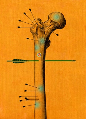
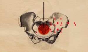 Illustration: Matthew Richardson


